lv mass index 2d Normal values for aorta in 2D echocardiography. Adjustment refers to adjusting for body surface area (BSA). Aorta, predicted. Aorta, standardized. Aortic valve regurgitation. Aortic valve stenosis. Left and right ventricle. Visual assessment . Grohe 34270EN1 Brushed Nickel Concetto Single Handle Single Hole Bathroom Faucet with SilkMove Ceramic Disc Cartridge - Free Metal Drain Assembly with purchase - FaucetDirect.com. The selected finish has been discontinued and is no longer available. Grohe 34 270. Item # bci1228174. The Grohe 34270EN1 has been discontinued.
0 · what is lv mass 2d
1 · lv stroke volume 2d teich
2 · lv mass index chart
3 · lv mass index 2d high
4 · lv mass and index calculator
5 · lv mass 2d meaning
6 · lv mass 2d abnormal
7 · left ventricular mass index chart
All rights reserved © 2016 Novikontas
Normal values for aorta in 2D echocardiography. Adjustment refers to adjusting for body surface area (BSA). Aorta, predicted. Aorta, standardized. Aortic valve regurgitation. Aortic valve stenosis. Left and right ventricle. Visual assessment . Our LV calculator allows you to painlessly evaluate the left ventricular mass, left ventricular mass index (LVMI for the heart), and the relative wall thickness (RWT). Read on and discover all the details of our LV mass .
Normal LV mass and LV mass index values established using linear, 2D, and 3D techniques stratified by sex are shown in Table 2. These numbers were derived from the entire cohort of 1,854 study subjects. Left ventricular mass (LVM) is a well-established measure that can independently predict adverse cardiovascular events and premature death. 1-3 Population-based studies have revealed that increased LVM and left .This LV mass index calculator helps diagnose the type of cardiac hypertrophy based on LVMI and relative wall thickness.
The data presented by Vasan et al 1 have clear and important implications in the way echocardiographic data should be reported and understood in routine practice because they make it possible to readily identify (hypertensive) .LVM and RWT. LVM is the acronym for Left Ventricular Mass. LV mass (LVM) is a vital prognostic measurement we obtain with echocardiography to manage hypertension. RWT is the acronym for Relative Wall Thickness and is an . Increased left ventricular mass (LVM) is a strong independent predictor for adverse cardiovascular events 1, 2, 3, and associated with impaired left ventricular (LV) . Verdecchia and colleagues studying 694 patients with body surface area indexed LV mass lower than 125 g/m 2, without additional adjustment for obesity and other metabolic .
In this study of LV mass determined by two-dimensional-guided M-mode echocardiography in a healthy population, the investigators confirm that (as before) gender, blood pressure, and body . Introduction. Normal values of left ventricular mass (LV M) and cardiac chamber sizes in asymptomatic subjects are prerequisites for the diagnosis of individuals with heart disease.Left ventricular hypertrophy is an independent risk factor for cardiovascular morbidity and sudden cardiac death. 1, 2 In addition, enlarged left ventricular, right ventricular, or left atrial .
Left ventricular (LV) hypertrophy is an independent cardiovascular risk factor associated with significant excess and morbidity and mortality rates. 1–3 There is now evidence for the effectiveness of antihypertensive agents, .where IVS = thickness of interventricular septum, LVEDD = LV end diastolic dimension, and PWT = posterior wall thickness. Indexed for body surface area, an LV mass index greater than 131 g/m 2 for men and greater than 100 g/m 2 for women represents LVH. 15 Other cutpoints for LVH have also been suggested, indexing for height raised to a power. 17–19 LVH is one of the . Left ventricular hypertrophy is an important risk factor in cardiovascular disease and echocardiography has been widely used for diagnosis. Although an adequate methodologic standardization exists currently, differences in measurement and interpreting data is present in most of the older clinical studies. Variability in border limits criteria, left ventricular mass .
Left ventricular (LV) hypertrophy is a strong marker of risk for future cardiovascular events.1 Two-dimensional echocardiographic measurement of LV wall thickness in combination with geometric formulas to calculate and convert myocardial volume to myocardial mass provides a reproducible, quantitative, noninvasive assessment of LV mass that is more accurate than . 2D Method: LV mass, g 66–150 151–171 172–182 >193 96–200 201–227 228–254 >255 LV mass/BSA, g/m 2 44–88 89–100 101–112 ≥113 50–102 103–116 117–130 ≥131 BSA, body surface area; LV, left ventricular. Bold values: Recommended and best validated. Click here for calculation of LV mass.

Assessment of LV mass was based on echocardiography in a sample of 905 middle‐aged men representative of the general population (aged 42 to 61 years). During the follow‐up period of 20 years, there were a total of 63 sudden cardiac deaths. In a comparison of the top versus the bottom quartile of LV mass adjusted by body surface area (>120 versus . Left ventricular hypertrophy (LVH), defined by the left ventricular mass index (LVMI), is highly prevalent in hemodialysis patients and a strong independent predictor of cardiovascular events. Compared to cardiac magnetic resonance imaging (CMR), echocardiography tends to overestimate the LVMI. Here, we evaluate the diagnostic . Calculated LV mass index cutoffs for concentric remodeling versus concentric hypertrophy or for normal geometry versus eccentric hypertrophy were 87 g/m 2 for women and 102 . a large meta-analysis of >22,000 healthy subjects, which determined LV mass using M-mode, 2D linear, or 2D area-length methods and divided subjects by age, sex, and .
what is lv mass 2d
Background— Left ventricular (LV) mass is an important predictor of morbidity and mortality, especially in patients with systemic hypertension. However, the accuracy of 2D echocardiographic LV mass measurements is limited because acquiring anatomically correct apical views is often difficult. We tested the hypothesis that LV mass could be measured more . As a tool for the assessment of LVH, the echocardiogram has been shown to be more sensitive than the ECG when both were compared with necropsy criteria for anatomic LVH. 9 Echocardiographically determined LV .
michael kors packable coat
Left ventricular (LV) mass (LVM) is a predictor of cardiovascular morbidity and mortality and commonly calculated using 1-dimensional (1D) echocardiographic methods. . 2D Measurements and Mass Calculation. . The reliability and validity of echocardiographic measurement of left ventricular mass index in hemodialysis patients. Nephron, 65 (2 .Figure 1. Calculation of left ventricular mass. mass LV = 1.05 (mass total – mass cavity) LV = left ventricle; 1.05 = mycoardial mass constant. Left ventricular hypertrophy (LVH) A diagnosis of left ventricular hypertrophy is based on total .
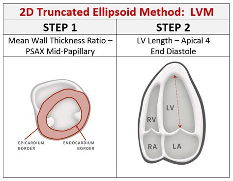
LVM calculated using 2D methods, truncated ellipsoid and area-length, is less affected by fluctuations in fluid and LVV, in contrast to 1D methods. . Rationale & objective: Left ventricular (LV) mass (LVM) is a predictor of cardiovascular morbidity and mortality and commonly calculated using 1-dimensional (1D) echocardiographic methods. These .The final recommendations from the ASE for LVM states that in normally shaped left ventricle, both M-mode and 2D echocardiographic formulas to calculate LV mass can be used. Normal values for these techniques should be reported indexed to BSA. Reference upper limits of normal LV mass by linear measurements are 95 g/m2 in women and 115 g/m2 in men. Assessment of LV mass was based on echocardiography in a sample of 905 middle‐aged men representative of the general population (aged 42 to 61 years). During the follow‐up period of 20 years, there were a total of 63 sudden cardiac deaths. In a comparison of the top versus the bottom quartile of LV mass adjusted by body surface area (>120 versus . Left ventricular mass index (LVMI) is a parameter used in echocardiography and cardiac MRI. LVMI is calculated using the following equations: LVMI = LVM (left ventricular mass)/body surface area; left ventricular mass = 0.8{1.04[([LVEDD + IVSd +PWd] 3 - LVEDD 3)]} + 0.6; where. LVEDD = LV end-diastolic dimension (mm)
Accurate characterization of left ventricular (LV) hypertrophy (LVH) is important since an increased LV mass (LVM) due to various forms of hypertrophy and remodeling is both a risk factor for . 2D Measurements and Mass Calculation. Tracing of the LV total area and LV cavity area was performed in the parasternal short-axis view at the level of the papillary muscle or just below the papillary muscle, with the papillary muscle considered part of the LV cavity. The tracer was positioned at the blood-tissue interface.
lv stroke volume 2d teich

lv mass index chart
Compensa Life Vienna Insurance Group SE. Compensa Life Vienna Insurance Group SE darbojas jau kopš 1993. gada un ir viena no vadošajām dzīvības apdrošināšanas sabiedrībām Baltijas valstīs.Compensa Life Vienna Insurance Group SE uzsāka savu darbību 1993.gadā Igaunijā, kā Seesam Elukindlustuse AS.. Compensa Life Vienna .
lv mass index 2d|what is lv mass 2d


