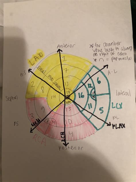
lv walls echo Learn how to identify and name the 17 segments of the left ventricle using echocardiography and other imaging modalities. The web page explains the standardized segmentation model, the . You can see you have UUID for the lvol you mentioned and along with it also sourced its filesystem type which is ext4. How to add UUID entry in /etc/fstab. Lets add this UUID entry in /etc/fstab using format –. So our entry will look like –.
0 · wall segments echo printable charts
1 · wall motion chart echo
2 · wall motion abnormalities echo
3 · lvh echo guidelines
4 · lvh echo criteria wall thickness
5 · lv wall thickness echo measurement
6 · lv wall thickness echo
7 · lv function assessment by echo
GAP, which stands for guaranteed asset protection, is optional insurance you can buy when buying a car. It helps cover the gap between what you owe on your auto loan and your car’s actual.
wall segments echo printable charts
Learn how to identify and name the 17 segments of the left ventricle using echocardiography and other imaging modalities. The web page explains the standardized segmentation model, the .Ejection fraction is the fraction of the end-diastolic volume (EDV, i.e blood volume in the ventricle at the end of diastole) that is pumped out during systole. Currently, two-dimensiona.Electronic calipers should be positioned on the interface between myocardial wall and cavity, and the interface between wall and pericardium. Perform at end-diastole (previously defined) .
Assessment of left ventricular systolic function has a central role in the evaluation of cardiac disease. Accurate assessment is essential to guide management and prognosis. Numerous echocardiographic techniques are used in the .
Ejection fraction is the fraction of the end-diastolic volume (EDV, i.e blood volume in the ventricle at the end of diastole) that is pumped out during systole. Currently, two-dimensional (2D) echocardiography for calculation of ejection .
LV Function and Haemodynamic Assessment Echocardiography. SYSTOLIC FUNCTION. Global Function. stroke volume: end-diastolic volume – end-systolic volume. cardiac output: Q = SV X HR. = (Aortic Area x V x Tej) x . The first and most commonly used echocardiography method of LVM estimation is the linear method, which uses end-diastolic linear measurements of the interventricular septum (IVSd), LV inferolateral wall .Echo Red Flags: When to suspect LVAD thrombosis Signs of LVAD Dysfunction: •Right‐shift of the IVS and LV enlargement •AoValve opening with every beat (9‐10/10 beats) •Blunted flow .
Identification and classification of left ventricular (LV) regional wall motion (RWM) abnormalities on echocardiograms has fundamental clinical importance for various cardiovascular disease.
Qualitative estimation of myocardial perfusion contrast echo is inferior to contrast enhanced 2D echocardiography with regard to visibility of all LV segments and appears slightly inferior with . Calculation of the left ventricular wall motion score index (WMSI) with transthoracic echocardiography allows the semi-quantification of left ventricular ejection .
Echocardiography is, therefore, an excellent screening tool for CA. Suggested echo indices to discriminate CA from other causes of LV hypertrophy comprise conventional LV remodeling and diastolic function assessment 6,7 and different deformation-based parameters (), such as the ratio of regional longitudinal strain values (relative apical sparing or septal apical . Calculation of the left ventricular wall motion score index (WMSI) with transthoracic echocardiography allows the semi-quantification of left ventricular ejection fraction ().Calculation of the LVEF with a WMSI demonstrates stronger agreement with measures obtained by cardiac MRI, the gold standard, than the traditional Simpson's biplane method. . Although attempts to standardize the terminology applied to the LV walls have been reported, 19,20 differences persist among the terms used by anatomists, pathologists, electrocardiographists, cardiac imagers, and .Systolic LV function: Regional wall motion | 17-segment model | Examples. Guidelines and Standards Recommendations for Cardiac ChamberQuantification by Echocardiography in Adults, 2015
Although attempts to standardize the terminology applied to the LV walls have been reported, 19,20 differences persist among the terms used by anatomists, pathologists, electrocardiographists, cardiac imagers, and clinicians. However, the pathologist’s view of infarcted myocardium lacks insights into the in vivo positioning of the LV walls. Left ventricular wall motion abnormalities are regularly assessed visually on echocardiography and cardiac MRI. The evaluation is primarily based on systolic wall thickening 1,2 and as a second criterion the systolic excursion. The localization of segmental wall motion abnormalities is done according to the cardiac segmentation model 6,7.

wall motion chart echo
Echo Assessment of LV Function: What Are We Really Trying to Measure) Jonathan R. Lindner, M.D. . Myocyte thickening: 8% Wall thickening: 40% Ejection Fraction: 60-70% 3 Young AA, et al., J Microsc 1998;191:131 Myocardial Sheet Composition 4. 1/19/20 3 Buckberg G, JTCS 2002:863 e R e C e l Streeter DD, et al. Circ Res 1969;24:339 Fiber .The segments close the the base of the heart, the basal segments are named for the wall to which they belong. There are six segments, numbered 1 thru 6, starting with the basal anteroseptal segment(1) and, going in a counter-clockwise direction, passing thru the basal anterior segment(2), basal anterolateral segment(3), basal inferolateral segment(4), basal . LV indicates left ventricle; HR, heart rate; bpm, beats per minute; LVID d, diastolic LV internal dimension; VST d, diastolic ventricular septal thickness; and LVPW d, diastolic LV posterior wall thickness. LV mass on echocardiography (Echo LV Mass) was calculated by using an uncorrected cube assumption from the formula 27 described in . Second, the LV wall thickness of the myocardial segments was obtained from the SAX views. Several studies evaluating the comparison of LV wall thickness measured by different imaging modalities emphasize the need for adequate standardization of image planes . For cardiac MRI and echocardiography, three-chamber measurements have better .
Introduction. Ventricular arrhythmia (VA) and sudden cardiac death (SCD) remain important problems in patients with heart failure (HF), despite the reduction in VA-associated deaths attributable to implantable cardioverter-defibrillators (ICDs) .The MADIT-CRT (Multicenter Automatic Defibrillator Implantation Trial With Cardiac Resynchronization Therapy) study .
Normal values for wall thickness are provided for middle-aged and older subjects. . the normal myocardial thickness values for SSFP are smaller than values established with the older spoiled gradient echo magnetic resonance imaging technique. LV mass obtained on SSFP images has been reported to be ≈20% smaller compared with gradient echo . Although transthoracic echocardiography remains the cornerstone of diagnostic cardiac ultrasound, transesophageal echocardiography (TEE) is a valuable complementary tool. . LV wall thickness can also be measured in these views at end-diastole to detect LV hypertrophy. To continue reading this article, you must sign in with your personal . Background—We sought to compare maximal left ventricular (LV) wall thickness (WT) measurements as obtained by routine clinical practice between echocardiography and cardiac magnetic resonance (CMR) and document causes of discrepancy. Methods and Results—One-hundred and ninety-five patients with hypertrophic cardiomyopathy (median . The authors found that 3D echocardiography was feasible and reproducible for the evaluation of LV size, shape, and function for patients with LV aneurysm. 3D echocardiography provided highly accurate quantification of .
ECHOCARDIOGRAPHY A. Cardiac Structure LV volumes and the pattern of hypertrophy can be well defined by echocardiography (Figure 1, Video 1 [ view video clip online], Table 1). Ventricular volumes in HCM are usually normal or slightly . wall thickness. LV hypertrophy, although usually asymmetric,can also be concen-tric. The distribution of .A Report from the American Society of Echocardiography Developed in Collaboration with the Society for Cardiovascular Magnetic Resonance. In Journal of the American Society of Echocardiography : official publication of the .
Second is the extent of involvement (ie, the number of myocardial segments involved) identified on echocardiography by the number of adjacent impaired myocardial segments. 7 Third is the percentage of LV wall that is replaced by fibrosis (scar thickness, indicating transmural infarction), 8, 9 which is identified on echocardiography by akinesis . The first and most commonly used echocardiography method of LVM estimation is the linear method, which uses end-diastolic linear measurements of the interventricular septum (IVSd), LV inferolateral wall thickness, and LV internal diameter derived from 2D-guided M-mode or direct 2D echocardiography. Echocardiographic measurements of minor axis and wall thickness and calculations from these two measurements of left ventricular end-diastolic volume and mass were performed in 24 patients and compared with angiocardiographic measurements of the same variables in corresponding patients. The echo-measured left ventricular end-diastolic chamber dimension .

intra-cardiac thrombus (especially LV) LV free wall rupture; Other advance echo techniques may be useful. DYNAMIC LVOT OBSTRUCTION WITHOUT ASYMMETRICAL SEPTAL HYPERTROPHY. dynamic LVOT obstruction with SAM can occur when ever a hyperdynamic state exists; can cause cardiogenic shock
Measurement of LV Size 3 1.1. Linear Measure-ments 3 1.2. Volumetric Measure-ments 3 1.3. Normal Reference . Regional Wall Motion during Infarction and Ischemia 11 3.4. Regional Abnormalities in the Absence of Cor- . Echocardiography (renamed the European Association of Cardiovascular Imaging [EACVI]) .
Currently, two-dimensional (2D) echocardiography for calculation of ejection fraction is the dominant method for assessing left ventricular function (systolic function). It should be noted, however, that three-dimensional (3D) echocardiography offers greater precision in the calculation of ejection fraction, and 3D measurements will presumably .Echocardiography plays a major role in the prognostic evaluation of hypertension (HTN). The main contribution of echo in the management of hypertension is the assessment of left ventricular mass (LVM). . Normal LV wall thickness; Increased LV cavity size; Increased LVM (Left ventricular mass) Decreased RWT (Relative Wall thickness) Concentric . LV mass and relative wall thickness. PLAX. 2D. LV wall thickness by itself does not define an individual as having left ventricular hypertrophy (LVH). Rather, the presence or absence of LVH is determined from LV mass after indexing to BSA. Wall thickness measurements, combined with the LV internal diameter in diastole, can be used to determine .speckle-tracking echocardiography. LV SYSTOLIC FUNCTION AND PROGNOSIS Assessment of LV systolic function is a cen-tral part of the evaluation of cardiac disease. . Cross-sectional view of the LV wall in the short-axis plane shows the longitudinal subendocardial myofiber orientation, trans-
Measurement of LV Size 3 1.1. Linear Measure-ments 3 1.2. Volumetric Measure-ments 3 1.3. Normal Reference . Regional Wall Motion during Infarction and Ischemia 11 3.4. Regional Abnormalities in the Absence of Cor- . Echocardiography (renamed the European Association of Cardiovascular Imaging [EACVI]) .
Inflict 1500 points of effect damage with a single effect activation in a Duel against Seto Kaiba at level 40 using Yami Yugi. UR Jewel x1 Achieve 18 win(s) with 1000 or less Life Points left in Duel World Duels.
lv walls echo|lv function assessment by echo


